Describe the Basic Structure of the Heart
The upper two chambers called atria and the lower two called the ventricles. The heart is a complex muscle that pumps blood through the three divisions of the circulatory system.

Basic Anatomy Of The Human Heart Cardiology Associates Of Michigan Michigan S Best Heart Doctors
The heart has a somewhat conical form and is enclosed by the pericardium.
. The coronary vessels that serve the heart pulmonary heart and lungs and systemic systems of the body. Structure of the heart. The heart is an organ about the size of your fist that pumps blood through your body.
In most people the heart is located on the left side of the chest beneath the breastbone. 1 layer is in direct contact with the heart itself and 1 year is in direct contact with the thoracic cavity. Chambers and Circulation through the Heart.
Heart Anatomy Location of the Heart. This tissue lines the inside of the heart and protects the valves and. The heart is composed of smooth muscle.
The ventricles pump the blood out of the heart. The Cardiac cycle is the. The atriu View the full answer.
The heart contains four inner chambers two on each side. One-way valves separate the four chambers. It comprises four chambers and several valves that regulate the normal flow.
Two atria and two ventricles. It is located in the middle cavity of the chest between the lungs. It is made up of multiple layers of tissue.
The inferior conical end of the heart is called the apex. It comprises four chambers. The muscular middle layer of the wall of.
It is positioned posteriorly to the body of the sternum with one-third situated on the right and two-thirds on the left of the midline. Structure of the Heart. Myocardium- This layer forms the heart muscles.
The heart is located posterior to the sternum left of the body midline between the lungs in the mediastinum. The heart is primarily made of a thick muscle layer called the myocardium surrounded by membranes. This is the muscular tissue of the heart.
The outer layer of the wall of the heart. The heart is divided into a right and left side by the septum. Heart Anatomy Heart Wall.
The heart measures 12 x 85 x 6 cm and weighs 310 g males and 255 g females Relations. In humans the heart is about the size of a clenched fist and it is divided into four chambers. It has four chambers which contract in a specific order allowing the human.
The heart of the bony fish has 2 chambers named atrium and ventricle. The posterosuperior surface of the heart is called the base. Your heart is at the center of your circulatory system.
There is an enlarged chamber preceeding the venous side of the heart and is known as sinus venosus. The circulatory system also called cardiovascular system is a vital organ system that delivers essential substances to all cells for basic functions to occur. Coronary circulation intrinsic to the heart takes blood directly from the main artery aorta coming from the heart.
4 chambers 2 atria and 2 ventricles that get blue deoxygenated blood from the body. Epicardium- This is a protective layer made of connective tissues. Cardiac conduction is the rate at which the heart conducts electrical impulses.
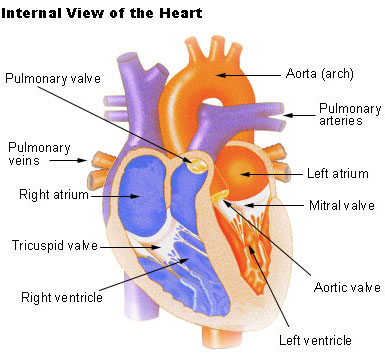
Brings deoxygenated blood from the body The right atrium receives it Passes through tricuspid valve to the right ventricle Deoxygenated blood leaves pulmonary artery Then reaches the lungs Pulmonary vein brings oxygenated blood from lungs Left atrium receives it Passes through bicuspid valve to the left ventricle Oxygenated. In this lab the class was divided into several groups and each group dissected either a sheep pig or cow heart. They pump out red oxygen-rich blood back to it.
The walls of the ventricles are relatively thicker than atrial walls. On the right side of the heart the right atrium and right ventricle work to pump oxygen-poor blood returning from the body back to the lungs to be reoxygenated. The shape of the heart is similar to a pinecone rather broad at the superior surface and.
Structure of the heart The heart can be found at the chests center underneath the sternum in a thoracic compartment. The human heart is located within the thoracic cavity medially between the lungs in the space. Your heart weighs about 10 - 12 ounces or 280 - 340 grams if you are a man and 8 -10 ounces or 230 - 280 grams if you are a woman.
Blood enters the heart at the upper chambers each called an atrium and when the atrial walls contract blood is pushed into the ventricles. The atria get blood coming back to the heart. In an adult the heart beats at an average of 60-80 times per minute.
The wall of the heart is made up of three layers. The basic structure of each heart is pretty much the same throughout each heart specimen. The left and right sides of the heart each have an upper chamber and a lower chamber.
The heart is a muscular organ that pumps blood throughout the body. The organs of the circulatory system are the heart arteries veins and capillaries. The structure of the heart The heart is a two-sided pump made up of four chambers.
The heart has four chambers two relatively small upper chambers called atria and two larger lower chambers called ventricles. Shape and Size of the Heart. The two atria are separated from each other by a thin muscular wall called the inter.
The heart is enclosed in a sac called the pericardium. The human heart is just roughly about the size of a fist. This system is a network of blood vessels such as arteries veins and capillaries that carries blood to and from all areas of your body.
After the dissection was complete each group moved around the room and collected data on all three different types of heart. The heart is made up of. Atria upper chambers Ventricles lower chambers Layers of heart.
Also commonly known as the cardiovascular system is a network composed of the heart as a centralised pump bloods vessels that distribute blood throughout the body and the blood. The hearts walls consist of three layers of tissue. The pericardium compartmentalizes and encloses the heart w its double layers.
Here are some more information about heart structure and function. The heart is a large muscular pump and is divided into two halves - the right-hand side and the left-hand side. Vantricle is the largest chamber and is the most muscular.
Think of the hearts position like an upside down pyramid with the apex below the base.

Heart Structure Bioninja
Anatomy And Physiology Of The Heart Normal Function Of The Heart Cardiology Teaching Package Practice Learning Division Of Nursing The University Of Nottingham

The Heart Anatomy How It Works And More
Seer Training Structure Of The Heart


Comments
Post a Comment